
Microscopy Lab
 Welcome to the Microscopy Lab! The Microscopy lab offers tools for biological research as well as technical assistance and training. The lab has three major microscopes - a transmission electron microscope (TEM), a scanning electron Microscope (SEM), and a laser-scanning confocal microscope (LSCM) equipped with a multi-photon (MP) laser. The microscopy lab also has both compound and stereomicroscopes for bright field and fluorescence imaging. Ancillary equipment necessary for sample preparation is also available.
Microscopy lab users can request full technical guidance, or once trained, can use the equipment independently. The lab manager is accessible by email, phone, or text message on or off campus. Additionally, the equipment availability is posted on the calendar below. The lab manager updates the calendar in real time. All microscopy requests need to be emailed with a project application form (see link above) to the lab manager stantond2@ufl.edu at least 24 hours in advance.
Fees
(all charges are per hour)
|
Microscope |
University of Florida |
External Universities |
industry |
|
Birghtfield and Dissecting Microscopes |
$15 |
$25 |
$40 |
|
Confocal w/o Multiphoton |
$40 |
$60 |
$80 |
|
Confocal w/ Multiphoton |
$50 |
$70 |
$100 |
|
SEM |
$60 |
$80 |
$120 |
|
TEM |
$65 |
$90 |
$130 |
|
Leica Microtome and Cryostat |
$15 |
$25 |
$40 |
|
Ultramicrotomes |
$30 |
$40 |
$50 |
|
Critical Point Drier |
$30 |
$40 |
$50 |
|
Sputter Coater |
$30 |
$40 |
$50 |
Equipment
Scanning Electron Microscope
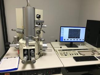
The center’s old Hitachi S-530 scanning electron microscope (SEM) was replaced with a newer model in the fall of 2015. A Hitachi S-4000 cold-field emission SEM was relocated from a University of Florida Lab in Gainesville and moved to the CREC and has been utilized frequently. This newer model allows us to have superior resolution over our previous model. The SEM allows for high resolution topography scans at high magnifications. It has a variety of uses from identifying morphological differences in mites, to identifying microbial pathogens such as bacteria and fungi on a leaf surface.
Transmission Electron Microscope
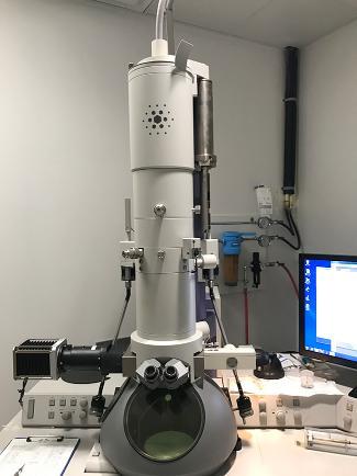
Our FEI Morgagni transmission electron microscope (TEM) was purchased in 2004. We upgraded our digital camera to an AMT XR50 5MP CCD camera in the spring of 2017 to increase image quality. The TEM is useful for ultrastructural studies. At the CREC, we predominately use our TEM to study the ultrastructure of various citrus tissue and identify morphological changes as well as pathogens. We also use the TEM in plant-vector interactions. Using the TEM allows us to identify the location of a pathogen in the insect and support transmission hypotheses with morphological studies.
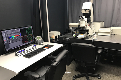
Confocal Laser-Scanning Microscope
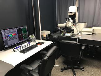
Our Leica SP8 Multi-photon confocal is our newest microscope. This microscope was purchased and installed in Spring of 2016. The confocal uses lasers to illuminate natural auto-fluorescence or detect fluorescing antibodies in a sample. The multi-photon laser can be tuned to excite different molecules at different wavelengths and can often penetrate deeper into tissue than the other lasers used in standard CLSM. Confocal laser-scanning microscopy (CLSM) has advanced our knowledge of cellular interactions. We can use CLSM to study protein-protein interactions, locate expressed genes using In situ hybridization and can even get three-dimensional renderings of organelles, cells, and tissues. The confocal eliminates light above and below the plane of focus to increase the resolution over traditional epi-fluorescence.
Epi-Florescence Microscope
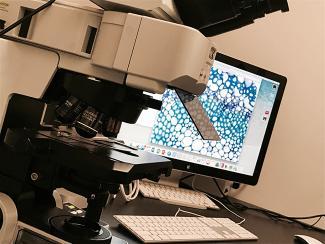
The CREC acquired a used Olympus BX61 in 2015. This upright, binocular microscope has a seven-position nose-piece with superior plan Apo objectives and a six-cube turret for fluorescent dichroic cubes. This microscope allows us to see natural fluorescence (auto-fluorescence) or antibody labeled proteins, organelles, or structures. Unlike the confocal, the fluorescent cubes cannot be ‘tuned’ and have a fix emission and excitation range for light to pass, thus a different cube or a cube with multiple filters are needed to see different wavelengths of light.
NightSea Stereomicroscope Adaptor
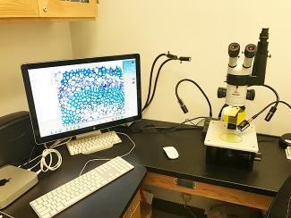
Our Leica Wild microscope was adapted in 2016 to function as a brightfield and fluorescence microscope by adding a NightSea system. This system allows for interchangeable light filters and detection filters for specific wavelengths. This has allowed us to adapt our fluorescence studies on a larger scale for genetic screening, compound screening, and for pathogen screening.
Staff
Daniel Stanton
Microscopy Lab Manager
863.956.8848
stantond2@ufl.edu